CIDPUSA.org Autoimmune diseases
MRI spine Child & CIDP
CIDP MRI in Children
Chronic inflammatory demyelinating polyneuropathy in a child:
clinical-spinal MR imaging correlation.
Likasitwattanakul S, Visrutaratna P.
Department of Pediatrics, Faculty of Medicine, Chiang Mai
University, Chiang Mai 50200, Thailand.
Spinal magnetic resonance (MR) imaging of a 3-year-old girl with
chronic inflammatory demyelinating polyneuropathy (CIDP) showed
thickened and marked enhancement of the lumbosacral nerve roots.
These abnormalities resolved after steroid treatment. MR imaging of
the cauda equina may be helpful in the diagnosis of CIDP.
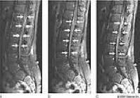
Figure 7 :Chronic inflammatory demyelinating polyneuropathy (CIDP) in a 13-year-old boy with peripheral neuropathy and gait disturbance. Sagittal fat-saturated T1-weighted images off the midline to the right (A), at the midline (B), and off the midline to the left (C). Chronic inflammatory demyelinating polyneuropathy (CIDP) in a 13-year-old boy with peripheral neuropathy and gait disturbance. D, E, F, and G, Axial postcontrast T1-weighted fat-saturated images through the lower thoracolumbar junction (D), conus medullaris (E), and nerve roots (F, G). The images show marked enhancement of the spinal nerve roots (arrows in A through F), with expansion of the nerve roots seen as they exit through the neural foramina (asterisks in F and G). This enlargement is typical of CIDP.
continued to cidp-studies page Chronic inflammatory demyelinating polyneuropathy
Links:
CIDPUSA.ORG
B-12 deficiency
- Small Fiber neuropathy
- neurological effects of CIDP
- Nerve Fiber types
- CIDP-EMG
- Multi Focal Motor neuropathy
- Lewis Summer
- Tips for CIDP
- Plasmapheresis
- CIDP-INFO
- CIDP-Rituxan
- CIDP-GBS-Handbook
- CIDP-family issue
- CIDP-Cyclosporin
- Polyneuropathy
- CIDP-GBS children
- Peripheral Neuropathy
- ALS & CIDP
- alcoholic poly neuropathy
- IVIg, Home to IVIg