Neuro Muscular Transmission disease
Myopathy Anterior Horn cell disease
😏cidpusa.org
WEAKNESS; Myopathy, Anterior horn cell disease, Neuropathies, Neuromuscular transmission disease
The anterior horn cells are somatotopically organized in the spinal cord. That is, medially (CENTER)located anterior horn cells innervate the proximal (shoulder & Hip) muscles, while laterally located ventral horn cells innervate more distal (hand & foot) muscles. The arrangement at cervical segments is shown in figure 2. This organization means that diseases that destroy anterior horn cells can result in highly selective weakness. Not only may a single muscle become weak, but only portions of the muscle may be affected. As a rule however the adjacent anterior horn cells will also be affected with weakness of adjacent muscles.
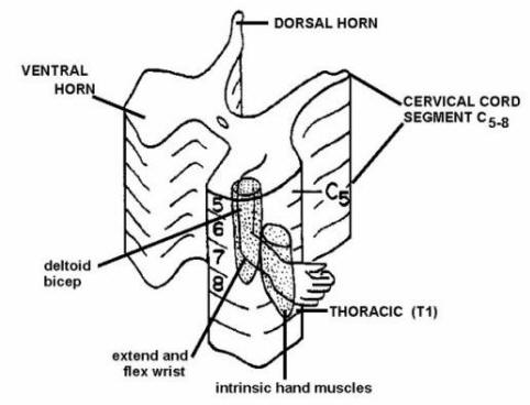
Figure 2 arrangement of anterior horn cells at cervical and the first thoracic levels. Because the anterior horn cells that innervate different muscles in the upper and lower extremities are present at different segments of the spinal cord, a whole extremity is not presented at a single level.
A note on the classification of dorsal and ventral root fibers.
The axons in the dorsal roots have been classified based upon their conduction velocities and their sizes. This has led to some confusion in the literature (and for medical students!!). The classifications scheme based upon fiber size uses Roman numerals. Thus, there are I, II, III and IV fiber types. You already have heard about the Ia fibers and that they are associated with muscle spindles and are large and fast conducting. You also have heard that the Ib fibers are associated with the Golgi tendon organs and are little smaller and slower conducting than the Ias. Also remember that II fibers are associated with muscle spindles but are slower conducting and smaller that the Ias and Ibs. II fibers are also associated with receptors carrying information from encapsulated endings used in two point discrimination, vibration and conscious proprioception. III fibers are smaller than Is and IIs and are only lightly myelinated and relatively slow conducting. Such fibers are associated with cooling and first pain. Finally, IV fibers are unmyelinated and convey second pain and warming.
Now lets turn to the classification that uses letters versus Roman numerals. The largest and fastest conducting fibers are called A fibers. Aa(alpha) fibers are comparable to the Ias and Ibs. Ab(alpha-beta) fibers are equivalent to II fibers in size and conduction velocities. Ad (deltas) are equivalent to IIIs and associated with cooling and first pain B fibers are smaller than A fibers, are lightly myelinated and are visceral afferents; they have no equivalent in the Roman numeral system. Finally, C fibers are unmyelinated and equivalent to IV fibers. In addition to carrying second pain and warming such fibers are postganglionic autonomics (but these do not travel in the dorsal roots).
What about ventral root fibers. The processes of lower motor neurons that innervate extrafusal muscle fibers are Aas (or just alpha motor neurons). The preganglionic autonomic axons in the ventral root are B fibers. Finally, there are axons in the ventral roots that innervate the intrafusal (not extrafusal) fibers of the muscle spindles. These are called Ag (gamma) motor neurons (no equivalent in Roman numerals).
Remember, A and B fibers are myelinated and Cs are not. In the Roman numeral system, just remember that only the IVs are not myelinated. This is important, since demyelinating diseases would affect the somatic and visceral afferents and efferent fibers in peripheral nerves, pain and temperature would not be affected.
Please continue to next page Myelinated Nerve Fiber & Muscle
😏