Sarcoidosis
Sarcoid is autoimmune
😏cidpusa.org
View larger version:Fig
Case 3. MRI of thighs at presentation. (A) T2‐weighted image demonstrating increased signal intensity in all muscle groups, particularly in vastus lateralis, medialis and intermedius. This is in keeping with widespread oedema. (B) T1‐weighted image in which the muscles are of normal signal intensity, because oedema (water) is iso‐intense with muscle. (C) On inversion recovery images widespread increased signal within the muscles is indicative of oedema. There is also increased signal in the subcutaneous fat in keeping with oedema at this site.
 Fig4.
Fig4.
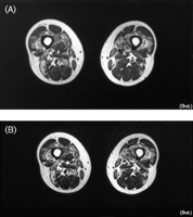
Case 3. (A) T1‐weighted and (B) T2‐weighted MRI demonstrating high signal intensity in vastus medialis and intermedius and the posterior aspect of vastus lateralis. High signal intensity is also seen in the short head of biceps femoris and adductor magnus. High signal change on both T1‐ and T2‐weighted images is characteristic of fatty infiltration. Mild atrophy is also noted.
Case 4
Diagnosis: Sarcoidosis—acute myositis, nodular myositis and interstitial lung disease.
Treatment: Prednisolone, azathioprine.
A 27 yr old professional guitarist presented with swelling, tightness and weakness of the forearms and pectoral muscles of 1‐month duration, and some shortness of breath for a week. Examination revealed swelling and thickening of the brachioradialis and pectoralis muscles bilaterally and of the right extensor digitorum brevis. There was no muscle weakness and reflexes and sensory examination were normal. In the chest there were fine inspiratory crepitations at both bases. There was no rash or lymphadenopathy.
The full blood count was normal, serum CK 532 U/l, angiotensin‐converting enzyme (ACE) 105 IU/l (normal range: 10–75), ESR 13 mm/h, ANA and Jo‐1 negative. Chest radiography showed bulky hilar shadowing and reticular shadowing at the lung bases. An EMG showed myopathic potentials, and an MRI of forearm muscles showed multiple regions of increased signal intensity on inversion recovery images suggestive of inflammation (Fig. 5⇔). Muscle biopsy of right brachioradialis revealed an interstitial inflammatory infiltrate of lymphocytes, histiocytes and multinucleated giant cells, with some muscle fibre degeneration and regeneration, but no vasculitis (Fig. 6⇔). These features were all in keeping with a diagnosis of acute sarcoid myositis with lung involvement.
The patient was treated with prednisolone and azathioprine. Within a few weeks his pain, swelling and muscle tenderness resolved and he was able to restart work. His shortness of breath improved. The dose of prednisolone was slowly reduced and azathioprine was increased. High‐resolution CT lung imaging showed considerable improvement and all of the blood tests normalized.
Four years later he developed two skin papules on his cheek and a palpable 2 cm nodule in the left triceps muscle without tenderness or weakness. Biopsy of the skin showed sarcoid‐like granulomatous inflammation. The clinical appearances of the triceps were of nodular myositis. Treatment with azathioprine was continued and he remains undereview.
😏
