CIDPUSA.ORG ❤
Autoimmune Diseases Web
Information on CARDIOMYOPATHY
alternatives treatment of autoimmune disease read our e-bookReturn to first page
A firm diagnosis typically requires a chest x ray to show whether the heart is
enlarged, an electrocardiogram to reveal any abnormal electrical activity of the
heart, and an echocardiogram, which uses sound waves to produce pictures of the
heart.
Other, more specific tests may also be needed. These include:
A radionuclide ventriculogram. This involves injecting low-dose radioactive
material (usually equal to that in a set of chest x rays) into a vein, through
which it flows to the heart. Pictures are generated by a special camera to show
how well the heart is functioning.
A cardiac catheterization. In this procedure, a thin plastic tube is inserted
through a blood vessel until it reaches the heart. A dye is injected and x rays
taken to assess the heart's structure and function.
Treatment
Since dilated cardiomyopathy is hard to diagnose early, it is rarely treated in
its beginning stage.
The goal of treatment is to relieve any complicating factor, if known, control
the symptoms, and stop the disease's progression. However, no cure now exists.
Therapy begins with the elimination of obvious risk factors, such as alcohol
consumption. Weight loss and dietary changes, especially salt restriction, may
also be advised.
Drugs used to treat the condition include:
Diuretics, which reduce excess fluid in the body;
Vasodilators, such as angiotensin-converting enzyme (ACE) inhibitors, which
relax blood vessels, helping to lower blood pressure and reducing the effort
needed by the heart to pump blood through the body;
Digitalis, which helps to improve pumping action and regulate heartbeat; and,
Calcium blockers or beta blockers, which may be used in some patients to help
regulate heartbeat and to alter the work of the heart muscle.
Also, patients with irregular heartbeats may be put on any of various drugs to
control the rhythm.
In critical cases where the condition is advanced and the patient does not
sufficiently respond to other treatments, a heart transplantation may be needed.
The patient's heart is replaced with a donor heart. Most heart transplant
recipients are under age 60 and in good health other than their diseased heart.
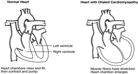
Course of the disease
As the heart enlarges, it steadily decreases its efficiency in pumping blood and
the amount of blood it can pump. As a result, some patients cannot perform even
simple physical activities.
However, the disease also may remain fairly stable for years, especially with
treatment and regular evaluation by a physician.
Unfortunately, by the time it is diagnosed, the disease often has reached an
advanced stage and heart failure has occurred. Consequently, about 50 percent of
patients with dilated cardiomyopathy live 5 years once heart failure is
diagnosed; about 25 percent live 10 years after such a diagnosis.
Typically, patients die from a continued decline in heart muscle strength, but
some die suddenly of irregular heartbeats.
For patients with advanced disease, heart transplantation greatly improves
survival: 75 percent of patients live 5 years after a transplantation. However,
in the United States, the scarcity of donor hearts limits the number of
transplantations to about 2,000 persons a year. Those who qualify for heart
transplantation often have to wait months, or even years, for a suitable donor
heart. Some patients with dilated cardiomyopathy die awaiting a transplant but,
according to recent studies, others improve enough from aggressive medical
treatment to be taken off the waiting list.
Also, some critically ill cardiomyopathy patients with declining heart function
use a small, implanted mechanical pump as a bridge to transplantation. Called
left ventricular assist devices (LVADs), these pumps take over part or virtually
all of the heart's blood pumping activity. The devices provided only temporary
assistance and are not now used as substitutes for heart transplantation.
Hypertrophic Cardiomyopathy
The second most common form of heart muscle disease is hypertrophic
cardiomyopathy. Physicians sometimes call it by other names: idiopathic
hypertrophic subaortic stenosis (IHSS), asymmetrical septal hypertrophy (ASH),
or hypertrophic obstructive cardiomyopathy (HOCM).
In hypertrophic cardiomyopathy, the growth and arrangement of muscle fibers are
abnormal, leading to thickened heart walls. The greatest thickening tends to
occur in the left ventricle (the heart's main pumping chamber), especially in
the septum, the wall that separates the left and right ventricles. The
thickening reduces the size of the pumping chamber and obstructs blood flow. It
also prevents the heart from properly relaxing between beats and so filling with
blood. Eventually, this limits the pumping action.