Autoimmune Muscle disease
Myositis is autoimmune
😏cidpusa.org
 View larger version:Fig2.
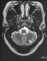
Case 2. T2 weighted MR image of brain showing high signal in the left cerebellar hemisphere in keeping with an area of ischaemia (arrow).Case 3
Fulminating onset polymyositis with myoglobinuric renal failure.
Treatment: Prednisolone, cyclophosphamide, i.v. Ig, cyclosporin, tracheostomy, PEG tube.
A 22 yr;old Caucasian man was admitted with a short history of fever, sweats, loss of weight, muscle pains and weakness. Before admission he developed shortness of breath, difficulty swallowing and noticed that he was passing dark urine. There was no history of drug or excess alcohol consumption. Examination revealed moderately severe proximal muscle weakness and tenderness but normal reflexes and sensation. The cardiovascular, respiratory and abdominal systems were normal.
Serum CK was >45 000 U/l, full blood count, urea and electrolytes, ESR and CRP were normal. Autoantibodies including ANA and Jo‐1 were negative. Urine contained myoglobin. EMG showed numerous, low‐amplitude, high‐frequency complex motor units with prominent fibrillation potentials and a full interference pattern on maximum volition indicating an active myopathic process. MRI showed extensive inflammatory change in the muscles of both thighs with relative sparing of semitendinosus, semimembranosus and biceps femoris muscles (Fig.3. The features were characteristic of fulminant polymyositis with myoglobinuria.
Serial lung function tests showed rapid deterioration and he required a tracheostomy and ventilation on the intensive therapy unit (ITU). The serum CK rose to 148 000 U/l and renal function deteriorated, peak serum urea 45.1 mmol/l, creatinine 277 Ámol/l, requiring haemofiltration to remove myoglobin from blood and preserve electrolyte balance. He was treated with i.v. methylprednisolone followed by oral prednisolone, bolus i.v. cyclophosphamide and i.v. Ig at 2 g/kg over 3 days. Progress was complicated by aspiration resulting in staphylococcal pneumonia with a lung abscess and pneumothorax. His muscle strength and bulk deteriorated considerably and he received a further course of i.v. Ig at 2 g/kg over 3 days. A gradual improvement in muscle strength followed, he was weaned off the ventilator but had persistent difficulty in swallowing and required a PEG tube. Renal function normalized, serum CK fell to 252 U/l and after 2 months he was transferred from ITU to the general wards. He had a third course of IVIG at 2 g/kg over 3 days and over the following month there was sufficient improvement in muscle strength, swallowing and respiratory function to enable the tracheostomy and PEG tubes to be removed.
Over the next year improvement in muscle strength continued with low‐dose oral prednisolone and azathioprine. The serum CK remained elevated between 200 and 600 U/l and renal function tests remained normal. Because of abdominal pain the azathioprine was changed to cyclosporin. He has remained well, back at full time work, and has no muscle weakness. Repeat MRI 18 months after the initial presentation shows no acute inflammation, but evidence of fatty change and mild atrophy
Rhabdomyolysis with myoglobinuria is a rare presentation of inflammatory muscle disease. However, in our series this has been present at disease onset in a total of four patients (5%). Precipitation of myoglobin in the nephron may rapidly lead to renal failure and therefore it is important to recognize a rising creatinine in the context of myoglobinuria. In this case haemofiltration was started very quickly in the ITU to preserve electrolyte balance and remove myoglobin. The serum potassium is extremely important in this situation because it may rise dramatically and lead to cardiac arrest.
In acute cases of inflammatory muscle disease, respiratory function must be monitored regularly as rapid deterioration can occur from respiratory muscle involvement or aspiration from oesophageal dysmotility. These patients should be admitted to the ITU early and treated aggressively to save life. Our patient made an excellent recovery.
This is another example of dysphagia responding to IVIg treatment but, as in case 1, requiring more than one course before the PEG tube could be removed. This case also illustrates our comments following case 1 regarding the ESR in inflammatory muscle diseases; in this patient it was normal at presentation.
continue to next page of polymyositis
😏