Synonyms
- Benign Pemphigus
- Old Age Pemphigus
- Parapemphigus
- Pemphigoid
- Senile Dermatitis Herpetiformis
Disorder Subdivisions
Related Disorders List
- Pemphigus
- Erythema Multiforme
- Pemphigoid, Benign Mucosal
- Dermatitis Herpetiformis
- Epidermolytic Hyperkerotosis
- Epidermolysis Bullosa
- Epidermolysis Bullosa Acquista
General Discussion
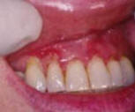
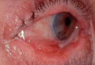
Bullous Pemphigoid is a rare, autoimmune, chronic skin disorder
characterized by blistering. This disorder occurs most frequently in elderly
people. Generalized blistering occurs in and under the upper layers of the
skin and usually subsides spontaneously within several months or years.
However, symptoms may recur. In some rare cases of Bullous Pemphigoid,
complications such as pneumonia may develop.
Symptoms
The first symptom of Bullous Pemphigoid is usually redness of the skin
surrounding a lesion, scar, and/or the navel. Within weeks, thin walled
blisters with clear fluid centers (bullae) appear on the undersurfaces of the
arms and legs (flexor surfaces), in the armpits (axillae), on the abdomen,
and/or around the groin. Unlike Pemphigus, Bullous Pemphigoid blisters usually
do not affect the mucous membrane lining the mouth; if they do they heal
rapidly.
The blisters are usually hard and tight, and contain clear or blood-tinged
fluid; they do not rupture easily. If the blisters do rupture, pain may occur
but healing is usually rapid.
Bullous Pemphigoid usually itches and in its early phase, itching and
hive-like patches may be the only symptoms.
After a few months, the symptoms of Bullous Pemphigoid often disappear
spontaneously, but they may recur for no apparent reason.
Causes
The exact cause of Bullous Pemphigoid is a autoimmune reaction by the body
against the glue holding the skin together; . Autoimmune disorders are
generated when the body’s natural defenses (e.g., the immune system with its
antibodies) against “foreign” or invading organisms, attack healthy tissue for
unknown reasons.
Certain drug reactions can produce skin lesions that are very similart to
Bullous Pemphigod. It is essential to determine whether side effects of the
pharmaceuticals may be causing the patient’s symptoms, or whether the blisters
are caused by Bullous Pemphigoid.
In 1990 it was determined that the gene for Bullous Pemphigoid is located on
chromosome 6 and has been mapped to 6p12-p11.
Affected Populations
Bullous Pemphigoid is a rare disorder that affects males and females in
equal numbers. This disorder primarily affect the elderly.
Related Disorders
Symptoms of the following disorders can be similar to those of Bullous
Pemphigoid. Comparisons may be useful for a differential diagnosis:
Pemphigus is an uncommon, sometimes life-threatening disease in which blisters
of varying sizes break out on the skin, the lining of the mouth, the vagina,
the thin covering of the penis and other mucous membranes (thin moist layers
that line the body's internal surfaces). The location and type of blister may
vary according to the type of Pemphigus. If left untreated, Pemphigus can be a
serious illness. Symptoms of this disorder include the appearance of soft
blisters in the outer layers of the skin especially on the neck, scalp, mucous
membranes, and/or the underarm and groin areas. (For more information on this
disorder, choose "Pemphigus" as your search term on the Rare Disease
Database.)
Erythema Multiforme is an allergenic, inflammatory skin disorder characterized
also by lesions on the skin and/or mucous membranes. The early symptoms of
this disorder may include red, elevated spots (erythematous macules or
papules) on the skin that may have fluid filled centers and that eventually
grow into larger blisters. Affected areas generally include the hands,
forearms, feet, and/or mucous membranes of the mouth, nose, and/or genitals.
The skin lesions and blisters caused by Erythema Multiforme generally appear
on both sides of the body and tend to heal in approximately 2 to 6 weeks.
Erythema Multiforme may also cause fever, joint pain, cough, and a sore
throat. (For more information on this disorder, choose "Erythema Multiforme"
as your search term in the Rare Disease Database.)
Benign Mucosal Pemphigoid is a rare chronic disease characterized by
blistering and scarring of the mucous membranes particularly in the mouth and
membranes that surround the eyes (conjunctiva). Initial symptoms include
redness and inflammation of these areas and scarring may occur on the
membranes of the eyes. Blisters may also develop in the mucous membranes of
the pharynx, esophagus, nose, urethra and/or vulva. (For more information on
this disorder, choose "Benign Mucosal Pemphigoid" as your search term on the
Rare Disease Database.
Dermatitis Herpetiformis is a rare chronic skin disorder that is characterized
by groups of severely itching blisters and elevated lesions. This disorder is
often associated with a sensitivity to foods that contain gluten
(gluten-sensitive enteropathy). The onset of Dermatitis Herpetiformis is
generally slow in adults; children may also be affected. Small, discrete
blisters and itchy smooth skin lesions that look like hives appear on the
head, elbows, knees, lower back, and/or buttocks. Itching and burning may be
almost intolerable, and the need to scratch may be overwhelming. (For more
information on this disorder, choose "Dermatitis Herpetiformis" as your search
term in the Rare Disease Database.)
Epidermolytic Hyperkeratosis (bullous type) is a rare hereditary skin disorder
characterized by the overgrowth of skin (hyperkeratosis) and abnormal redness
of the skin (erythroderma). The symptoms of this disorder are present at birth
and may range from mild to severe. The skin may appear to have warts or
blisters, and to be thick over most of the body, particularly in the skin
creases over joints. Epidermolytic Hyperkeratosis can be detected before birth
by amniocentesis (microscopic examination of the fluid that surrounds the
developing fetus). (For more information on this disorder, choose "Epidermolytic
Hyperkeratosis" as your search term in the Rare Disease Database.)
Epidermolysis Bullosa refers to a group of rare skin diseases characterized by
fragile skin, blisters, and small fluid-filled lesions that develop following
minor trauma to the skin. The mucous membranes are also involved in some forms
of Epidermolysis Bullosa. Healing may be impaired. Blisters may leave multiple
scars and/or damage the underlying muscle tissue. Most types of Epidermolysis
Bullosa are inherited, and they usually first appear during childhood. (For
more information on these disorders, choose "Epidermolysis Bullosa" as your
search term in the Rare Disease Database.)
Epidermolysis Bullosa Acquista is a rare autoimmune disorder of the skin that
typically affects middle-aged and elderly people. Trauma to the skin can cause
blisters on the elbows, knees, pelvis, buttocks, and/or scalp. Increased
levels of IgG (an immunoglobulin) are usually found around the blisters. After
the blisters heal, scars usually remain. (For more information on this
disorder, choose "Epidermolysis Bullosa Acquista" as your search term in the
Rare Disease Database.)
Standard Therapies
Corticosteroid drugs are given to people with Bullous Pemphigoid to help
reduce the number of blisters. The dosage of these immune suppressant drugs is
lower for people with Bullous Pemphigoid than the dosage prescribed to treat
Pemphigus. Corticosteroid drugs (usually prednisone) can be discontinued in
approximately 50 percent of cases of Bullous Pemphigoid because the patients
eventually go into remission. The remainder of patients may require
maintenance therapy. Because many people with Bullous Pemphigoid are elderly,
decisions about whether to treat with drugs that alter the immune system (such
as corticosteroids) must be individualized because it may make fragile
patients more susceptible to infections.
Investigational Therapies
Immunosuppressive drugs such as azathioprine and methotrexate, have been
used on an experimental basis in combination with corticosteroid drugs to
treat Bullous Pemphigoid.
Cyclosporine (Sandimmune) may be of potential benefit for treating a number of
autoimmune skin diseases including Pemphigus, Bullous Pemphigoid, Posterior
Uveitis, and Behcet's Syndrome. However, careful monitoring by a physician of
people taking cyclosporine is necessary to guard against possible toxic side
effects. Relapses of Bullous Pemphigoid can occur when the drug therapy is
stopped. More research is needed before cyclosporine can be recommended as a
treatment for all but the most severe cases of Bullous Pemphigoid.
Clinical trials are underway to study possible new therapies for Bullous
Pemphigoid. Interested persons may wish to have their physician contact:
W.R. Gammon, M.D.
Dept. of Dermatology
University of North Carolina
137 NC Memorial Hospital
Chapel Hill, NC 27514
References
McKusick VA., ed. Online Mendelian Inheritance in Man (OMIM). Baltimore.
MD: The Johns Hopkins University; Entry No: 113810; Last Update: 7/21/99.
TEXTBOOKS
Thoene JG., ed. Physicians’ Guide to Rare Diseases. Montvale, NJ: Dowden
Publishing Company Inc; 1995:651
Berkow R., ed. The Merck Manual-Home Edition. Whitehouse Station, NJ: Merck
Research Laboratories; 1997:968.
Beers MH, Berkow R., eds. The Merck Manual, 17th ed. Whitehouse Station, NJ:
Merck Research Laboratories; 1999:830.
Champion RH, et al., eds. Textbook of Dermatology. 5th ed. Cambridge, MA:
Blackwell Scientific Publications; 1992:1647-51.
Habif TP, ed. Clinical Dermatology. 2nd ed. St. Louis, MO: The C.V. Mosby
Company; 1990:415-16.
Bennett JC, Plum F., eds. Cecil Textbook of Medicine. 20th ed. Philadelphia,
PA: W.B. Saunders Co; 1996:2205-06.
REVIEW ARTICLES
Roeder C, et al., Psoriatic erythroderma and bullous pemphigoid treated
successfully with acitretin and azathioprine. Eur J Dermatol. 1999;9:537-39.
Nousari HC, et al., Pemphigus and bullous pemphigoid. Lancet. 1999;354:667-72.
Trueb RM, et al., Childhood bullous pemphigoid: report of a case with
characterization of the targeted antigens. J Am Acad Dermatol. 1999;40:338-44.
Scott JE, et al., The blistering diseases. Med Clin North Am. 1998;82:1239-83.
JOURNAL ARTICLES
Korman NJ., New and emerging therapies in the treatment of blistering
diseases. Dermatol Clin. 2000;105:113-23.
Kuboki T, et al., [Recurrent bleeding tendency due to factor VIII inhibitor
during therapy for bullous pemphigoid. Rinsho Ketsueki. 1999;40:1087-92.
Mainguy G, et al., Regulation of epidermal bullous pemphigoid antigen 1
(BPAG1) synthesis by homeoprotein transcription factors. J Invest Dermatol.
1999;113:643-50.
Kirtschig G, et al., IgA basement membrane zone autoantibodies in bullous
pemphigoid detect epidermal antigens of 270-280 kDa, 230 kDa, and 180 kDa
molecular weight by immunoblotting. Clin Exp Dermatol. 1999;24:302-07.
Egan CA, et al., The immunoglobulin A antibody response in clinical subsets of
mucous membrane pemphigoid. Dermatology. 1999;198:330-35.
Rhodes LE, et al., Blister fluid cytokines in cutaneous inflammatory bullous
disorders. Acta Derm Venereol. 1999;79:288-90.
Jung M, et al., Increased risk of bullous pemphigoid in male and very old
patients: A population-based study on incidence. J Am Acad Dermatol. 1999;41
(2 Pt 1):266-68.