| |
Dr Khan , CIDPUSA & Nanotech Medical Center have done
their research and published the
autoimmune diseases E-BOOK called
, "The Flame Within". Which contains treatment of alzheimers,
CIDP, neuropathies, Pemphigus, acne, alopecia , every type of
arthritis and many more by simple antibiotics. Save your money and
purchase this book online today 24 hour delivery to your email.
Check this offer on our home page. Just remember most of the
research reported in the news is supported by the large pharma
business. Our research was done at university medical centers and we
make no money on the recommended antibiotic treatments with
permanent cures.
 
Pemphigus is a group of rare autoimmune skin disorders characterized by the
development of blisters in the outer layer of the skin (epidermis) and mucous
membranes (thin moist layers that line the body's internal surfaces). The
location and type of blisters vary according to the type of Pemphigus. If left
untreated Pemphigus can be a serious illness. Mostly misdiagnosed
as Herpes.
If you have had chemical exposure, accidents, injury, surgery, arthritis,
then pemphigus and pemphigoid are more likely.

Symptoms
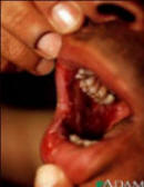
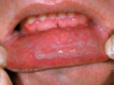
Blisters in the outer layer of the skin are common to all types of Pemphigus.
Blisters develop due to the destruction of the "cement" that holds cells
together (epidermal acantholysis) resulting in the separation of cells from one
another. Soft (flaccid) blisters generally occur on the neck, scalp, mucous
membranes, and/or underarm (axillary) and groin areas (inguinal). Most patients
with Pemphigus have deposits of IgG (an immune system antibody that defends
against foreign substances) around the blistered areas (in the epidermal cells
called keratinocytes). Antiepidermal antibodies directed against skin cells are
typically present in the fluid of the blisters. The diagnosis of Pemphigus
requires microscopic examination of cells in the blisters as well as detection
of the IgG antibodies that characterize this disease.
Pemphigus Vulgaris is the most common form and may begin with isolated blisters
on the scalp, and then in the mouth. These may persist for several months and
may be followed by blistering of the esophagus, nose, rectum, and/or the
membranes that line the inner surfaces of the eyelids (conjunctiva). The
blisters are soft; they break easily and heal poorly. Pressure on the borders of
blisters causes them to spread. Pressure on normal-looking skin can cause it to
blister (Nikolsky sign) in people with Pemphigus Vulgaris. If left untreated,
Pemphigus Vulgaris may cause life-threatening complications.
Pemphigus Vegetans is a variation of Pemphigus Vulgaris. The blisters are
fast-growing and have large (hypertrophic) lesions that are usually located in
the groin (inguinal) and armpit (axillary) areas.
Pemphigus Foliaceus is less severe and a less common form of the disorder. Soft
blisters typically occur close to the surface of the skin. When they break, they
ooze and become crusty, scaly, and susceptible to infection. Blisters may occur
on the scalp, face, upper chest, and back; the mucous membranes are usually not
affected. Small, horny plugs attached to the undersurface of the affected skin
also may be seen.
Another type of Pemphigus Foliaceus occurs in South America, particularly Brazil
and Colombia, and is called Fogo Selvagem.
When individuals have symptoms of both Pemphigus Foliaceus and Systemic Lupus
Erythematosus, they are said to have Pemphigus Erythematosus. Lupus, also known
as SLE, is an inflammatory disease of connective tissue. (For more information
on this disorder, choose "Lupus" as your search term in the Rare Disease
Database.)
Pemphigus may also occur as a result of an adverse reaction to certain drugs
such as d-penicillamine and rifampin; symptoms usually resemble those of
Pemphigus Foliaceus rather than Pemphigus Vulgaris. Some research suggests that
Pemphigus Herpetiformis is a subtle form of Pemphigus with its own
characteristic blisters. However, blisters that form during a relapse may
resemble those of Pemphigus Foliaceus.
In Benign Familial Pemphigus (Hailey-Hailey Disease), recurrent blisters are
seen primarily on the neck, groin, and armpits. Blisters may recur because of
sweating, skin infections, and exposure to extreme heat and/or ultraviolet
light.
Causes
Most forms of Pemphigus are generally considered to be autoimmune-related.
Autoimmune disorders are caused when the body's natural defenses (e.g.,
antibodies) against "foreign" or invading organisms begin to attack healthy
tissue for unknown reasons. Usually triggered by exposure to chemicals , dyes,
trauma, surgery and exposure to infections. Carpet Shampoo is well known to
trigger the blistering in Kawasaki disease.
Benign Familial Pemphigus (Hailey-Hailey disease) is inherited as an autosomal
dominant trait. Human traits, including the classic genetic diseases, are the
product of the interaction of two genes, one received from the father and one
from the mother. In dominant disorders, a single copy of the disease gene
(received from either the mother or father) will be expressed "dominating" the
other normal gene and resulting in the appearance of the disease. The risk of
transmitting the disorder from affected parent to offspring is 50 percent for
each pregnancy regardless of the sex of the resulting child.
( Pemphigus Foliaceus) is an autoimmune blistering
disorder that may be triggered by a substance transmitted through the bite of
blackflies.
Pemphigus may also occur following x-ray exposure or adverse reaction to drugs
such as d-penicillamine or rifampin.
Affected Populations
Pemphigus affects males and females in equal numbers and is most common in
middle-aged and elderly people. However, cases of children with Pemphigus have
been reported. This disorder has been found in all ethnic groups and races, but
is more common in people of Jewish or Mediterranean ancestry. Pemphigus occurs
once in 100,000 people in the United States.
Related Disorders
Symptoms of the following disorders can be similar to those of Pemphigus.
Comparisons may be useful for a differential diagnosis:
Bullous Pemphigoid is a chronic mild skin disorder that generally affects
elderly people. It is characterized by large firm fluid-filled blisters (bullous
pemphoid) that heal quickly and typically disappear in several months or years.
However they may recur later. Early symptoms include redness on the skin
followed within weeks by the appearance of blisters. The mucous membranes are
rarely affected by Bullous Pemphigoid. (For more information on this disorder,
choose "Bullous Pemphigoid" as your search term in the Rare Disease Database.)
Darier Disease (Darier-White Disease or Keratosis Follicularis) is a progressive
inherited skin disorder characterized by widespread firm elevated lesions on the
skin and mucous membranes; abnormal changes of the finger and toe nails may also
occur. Symptoms usually begin with a sensation of itching or burning on the
skin, especially the scalp, forehead, face, neck, and back. Firm, elevated spots
(papules) appear and typically become large and darkened; eventually these
papules become scaly and crusty. As these spots enlarge, they may come together
and form larger areas. The symptoms of Darier Disease tend to become more severe
during periods of emotional stress or with exposure to sunlight. (For more
information on this disorder, choose "Darier" as your search term on the Rare
Disease Database.)
Epidermolytic Hyperkeratosis (bullous type) is a rare hereditary skin disorder
characterized by the overgrowth of skin (hyperkeratosis) and an abnormal redness
of the skin (erythroderma). The symptoms are present at birth and may range from
mild to severe. The skin may appear "warty," blistered, and thick over most of
the body, particularly in the skin creases over joints. The disorder can be
detected before birth by amniocentesis (microscopic examination of the fluid
that surrounds the developing fetus). (For more information on this disorder,
choose "Epidermolytic Hyperkeratosis" as your search term in the Rare Disease
Database.)
Erythema Multiforme is an allergic inflammatory skin disorder characterized by
lesions that develop on the skin and mucous membranes. The early symptoms may
include red, elevated spots (erythematous macules or papules) that may have
fluid filled centers and eventually grow into larger blisters. Affected areas
generally include: hands, forearms, feet, and/or mucous membranes of the mouth,
nose and/or genitals. The skin lesions and blisters caused by Erythema
multiforme generally appear on both sides of the body and tend to heal in
approximately 2 to 6 weeks. Erythema Multiforme may also cause fever, joint
pain, cough, and a sore throat. (For more information on this disorder, choose "Erythema
Multiforme" as your search term in the Rare Disease Database.)
Epidermolysis Bullosa refers to a group of rare hereditary skin diseases
characterized by fragile skin; blisters and small fluid-filled lesions develop
following minor trauma. In some forms of Epidermolysis Bullosa, the mucous
membranes are involved. Healing may be impaired in some forms of this disorder
resulting in multiple scars and/or damage to underlying muscle tissue. (For more
information on these disorders, choose "Epidermolysis Bullosa" as your search
term in the Rare Disease Database.)
Epidermolysis Bullosa Acquista is a rare autoimmune disorder of the skin that
typically affects middle-aged and elderly people. Trauma may cause blisters on
the skin of the elbows, knees, pelvis, buttocks, and scalp. Increased levels of
IgG are usually found around the blisters; scars usually remain after healing.
(For more information on this disorder, choose "Epidermolysis Bullosa" as your
search term in the Rare Disease Database.)
Dermatitis Herpetiformis is a rare chronic skin disorder that is characterized
by groups of severely itching blisters and elevated lesions. This disorder is
often associated with a sensitivity to foods that contain gluten
(gluten-sensitive enteropathy). The onset of this disorder in generally slow in
adults, but children may also be affected. Small, discrete blisters and itchy
smooth lesions similar to hives may appear on the head, elbows, knees, lower
back, and buttocks. Itching and burning may be almost intolerable, and the need
to scratch may be overwhelming. (For more information on this disorder, choose
"Dermatitis Herpetiformis" as your search term in the Rare Disease Database.)
Standard Therapies
Corticosteroids are the most widely used drugs for treating Pemphigus.
Frequently, topical corticosteroid ointments can relieve inflammation and
itching, and oral (systemic) corticosteroids such as prednisone relieve
inflammation and suppress the immune system.
Immunosuppressive drugs such as cyclosporine, cyclophosphamide, azathioprine, or
methotrexate may be prescribed to treat severe cases of Pemphigus. Cytotoxic
drugs may also be used to suppress the immune system. Gold compounds such as
auranofin may be given to relieve inflammation and to attempt to suppress the
immune system (chrysotherapy). The drug Dapsone may also be prescribed but
should be used with extreme caution. To reduce immediate or long-term side
effects, drug therapy may be stopped temporarily or changed.
Antibiotic drugs or creams may be given to manage infection and relieve
inflammation. Silver sulfadiazine cream also may be used. Dusting the patient
and their bed sheets with talcum powder may relieve the discomfort of raw skin.
Genetic counseling may be beneficial for patients and their families if they
have the hereditary form of Pemphigus. Other treatment is symptomatic and
supportive.
Investigational Therapies
Plasmapheresis may benefit some people with Pemphigus. This procedure is a
method for removing unwanted substances (toxins, metabolic substances and plasma
parts) from the blood. Blood is removed from the patient and blood cells are
separated from plasma. The patient's plasma is then replaced with other human
plasma and the blood is retransfused into the patient. This therapy is still
under investigation to analyze side effects and long-term effectiveness. More
research is needed before plasmapheresis can be recommended for use in all but
the most severe cases of Pemphigus.
Researchers are investigating a new method of plasmapheresis for individuals
with Pemphigus. An immunoglobin-free albumin solution is substituted for the
plasma and blood that is usually transfused into an affected individual. This
procedure may decrease the autoimmune response of the antibodies in the patient.
Scientists are studying a new approach to treatment of Pemphigus that uses
extracorporeal (outside the body) photopheresis. In this process, the blood is
withdrawn, exposed to ultraviolet light, and then returned to an affected
individual. More study is needed to determine the long-term safety and
effectiveness of this procedure.
Surgery is also being investigated to treat Pemphigus when it is not responsive
to standard therapy. Surgery involves removing the blistered skin and applying
skin grafts.
Researchers at the National Institutes of Health (NIH) are conducting diagnostic
procedures such as immunofluorescence testings and immumoprecipitation studies.
More studies are needed to determine the long-term safety and effectiveness of
these techniques for the treatment of Pemphigus. For more information, contact:
Intravenous immunoglobulin therapy is being studied as a treatment for
individuals with Pemphigus. Initial research shows that this form of therapy may
be an effective alternative for individuals with who are unresponsive to
conventional therapy, dependent upon corticosteroids, or experience adverse side
effects to corticosteroids. More research is necessary to determine the
long-term safety and effectiveness of intravenous immunoglobulin therapy for
individuals with Pemphigus.
Please contact us for
help For any questions on treatment cause insurance problems we can help.
www.cidpusa.org
www.cidpusa.org/P/ivig.htm
http://www.cidpusa.org/disease.html
http://www.cidpusa.org/Lahore.html
|
|