INTRODUCTION ON HOW TO SAVE A MILLION DOLLARS In the treatment of
Cardiomyopathy
Cardiomyopathy is a disease of the heart muscle. The heart loses its ability
to pump blood and, in some instances, heart rhythm is disturbed, leading to
irregular heartbeats, or . Usually, the exact cause of the muscle damage is
never found.
Cardiomyopathy differs from many other heart disorders in a couple of ways.
First, the types not related to coronary atherosclerosis are fairly uncommon.
Cardiomyopathy affects about 50,000 Americans. However, the condition is a
leading reason for heart transplantation.
Second, unlike many other forms of heart disease that affect middle-aged and
older persons, certain types of cardiomopathies can, and often do, occur in the
young. The condition tends to be progressive and sometimes worsens fairly
quickly. Majority of the time the cause is autoimmune.

NONISCHEMIC CARDIOMYOPATHY
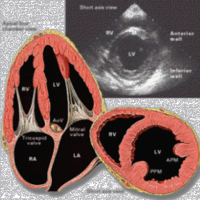
As noted, there are various types of cardiomyopathy. These fall into two
major categories: "ischemic" and "nonischemic" cardiomyopathy.
- Ischemic cardiomyopathy typically refers to heart muscle damage that
results from coronary artery disease, such as heart attack, and will not be
discussed here (see page 8 on how to get information on the disorder).
- Nonischemic cardiomyopathy includes several types. The three main types
are covered in this fact sheet. They are: dilated, hypertrophic, and
restrictive. The name of each describes the nature of its muscle damage.
Dilated (Congestive) Cardiomyopathy
By far the most common type of nonischemic cardiomyopathy, the dilated
(stretched) form occurs when disease-affected muscle fibers lead to enlargement,
or dilation, of one or more chambers of the heart. This weakens the heart's
pumping ability. The heart tries to cope with the pumping limitation by further
enlarging and stretching--a process known as "compensation."
Dilated cardiomyopathy occurs most often in middle-aged people and more often
in men than women. However, the disease has been diagnosed in people of all
ages, including children.
In most cases, the disease is bb--a specific cause for the damage is never
identified.
But some factors have been linked to the disease's occurrence. For instance,
alcohol has a direct suppressant effect on the heart. Dilated cardiomyopathy can
be caused by chronic, excessive consumption of alcohol, particularly in
combination with dietary deficiencies. Also, dilated cardiomyopathy occasionally
occurs as a complication of pregnancy and childbirth. Other factors are: various
infections, mostly viral, which lead to an inflammation of the heart muscle (myocarditis);
toxins (such as cobalt, once used in beers, for instance); and, rarely,
heredity.
Some drugs, used to treat a different medical condition, also can damage the
heart and produce dilated cardiomyopathy. Such drugs include doxorubicin and
daunorubicin, both used to treat cancer.
Whatever the cause, the clinical and pathological manifestations of dilated
cardiomyopathy are usually the same.
Symptoms
Dilated cardiomyopathy can be present for several years without causing
significant symptoms. With time, however, the enlarged heart gradually weakens.
This condition is commonly called "," and it is the hallmark of dilated
cardiomyopathy. Typical signs and symptoms of heart failure include: fatigue;
weakness; shortness of breath, sometimes severe and accompanied by a cough,
particularly with exertion or when lying down; and swelling of the legs and
feet, resulting from fluid accumulation that may also affect the lungs
(congestion) and other parts of the body. It also produces abnormal weight gain.
(The cough and congestion mimic and, therefore, can be misdiagnosed as pneumonia
or acute bronchitis. Also, heart failure is often from heart disease other than
cardiomyopathy.)
Because of the congestion, some physicians use the older term "congestive
cardiomyopathy" to refer to dilated cardiomyopathy. In advanced stages of the
disease, the congestion may cause pain in the chest or abdomen.
In advanced stages, some patients develop irregular heartbeats, which can be
serious and even life threatening.
Diagnosis
Once symptoms appear, the condition may be tentatively diagnosed based on a
physical examination and a patient's medical history. More often, though,
further examination is needed to differentiate dilated cardiomyopathy from other
causes of heart failure.
A firm diagnosis typically requires a chest x ray to show whether the heart
is enlarged, an to reveal any abnormal electrical activity of the heart,
and an echocardiogram, which uses sound waves to produce pictures of the heart.
Other, more specific tests may also be needed. These include:
- A radionuclide ventriculogram. This involves injecting low-dose
radioactive material (usually equal to that in a set of chest x rays) into a
vein, through which it flows to the heart. Pictures are generated by a special
camera to show how well the heart is functioning.
- A cardiac catheterization. In this procedure, a thin plastic tube is
inserted through a blood vessel until it reaches the heart. A dye is injected
and x rays taken to assess the heart's structure and function.
Treatment
Since dilated cardiomyopathy is hard to diagnose early, it is rarely treated
in its beginning stage.
The goal of treatment is to relieve any complicating factor, if known,
control the symptoms, and stop the disease's progression. However, no cure now
exists.
Therapy begins with the elimination of obvious risk factors, such as alcohol
consumption. Weight loss and dietary changes, especially salt restriction, may
also be advised.
Drugs used to treat the condition include:
- Diuretics, which reduce excess fluid in the body;
- Vasodilators, such as angiotensin-converting enzyme (ACE) inhibitors,
which relax blood vessels, helping to lower blood pressure and reducing the
effort needed by the heart to pump blood through the body;
- Digitalis, which helps to improve pumping action and regulate heartbeat;
and,
- Calcium blockers or beta blockers, which may be used in some patients to
help regulate heartbeat and to alter the work of the heart muscle.
- IVIG has been shown effective in several
studies.
Also, patients with irregular heartbeats may be put on any of various drugs
to control the rhythm.
In critical cases where the condition is advanced and the patient does not
sufficiently respond to other treatments, a heart transplantation may be needed.
The patient's heart is replaced with a donor heart. Most heart transplant
recipients are under age 60 and in good health other than their diseased heart.
Course of the disease
As the heart enlarges, it steadily decreases its efficiency in pumping blood
and the amount of blood it can pump. As a result, some patients cannot perform
even simple physical activities.
However, the disease also may remain fairly stable for years, especially with
treatment and regular evaluation by a physician.
Unfortunately, by the time it is diagnosed, the disease often has reached an
advanced stage and heart failure has occurred. Consequently, about 50 percent of
patients with dilated cardiomyopathy live 5 years once heart failure is
diagnosed; about 25 percent live 10 years after such a diagnosis.
Typically, patients die from a continued decline in heart muscle strength,
but some die suddenly of irregular heartbeats.
For patients with advanced disease, heart transplantation greatly improves
survival: 75 percent of patients live 5 years after a transplantation. However,
in the United States, the scarcity of donor hearts limits the number of
transplantations to about 2,000 persons a year. Those who qualify for heart
transplantation often have to wait months, or even years, for a suitable donor
heart. Some patients with dilated cardiomyopathy die awaiting a transplant but,
according to recent studies, others improve enough from aggressive medical
treatment to be taken off the waiting list.
Also, some critically ill cardiomyopathy patients with declining heart
function use a small, implanted mechanical pump as a bridge to transplantation.
Called left ventricular assist devices (LVADs), these pumps take over part or
virtually all of the heart's blood pumping activity. The devices provided only
temporary assistance and are not now used as substitutes for heart
transplantation.
Hypertrophic Cardiomyopathy
The second most common form of heart muscle disease is hypertrophic
cardiomyopathy. Physicians sometimes call it by other names: idiopathic
hypertrophic subaortic stenosis (IHSS), asymmetrical septal hypertrophy (ASH),
or hypertrophic obstructive cardiomyopathy (HOCM).
In hypertrophic cardiomyopathy, the growth and arrangement of muscle fibers
are abnormal, leading to thickened heart walls. The greatest thickening tends to
occur in the left (the heart's main pumping chamber), especially in the
septum, the wall that separates the left and right ventricles. The thickening
reduces the size of the pumping chamber and obstructs blood flow. It also
prevents the heart from properly relaxing between beats and so filling with
blood. Eventually, this limits the pumping action.
Hypertrophic cardiomyopathy is a rare disease, occurring in no more than 0.2
percent of the U.S. population. It can affect men and women of all ages.
Symptoms can appear in childhood or adulthood.
Most cases of hypertrophic cardiomyopathy are inherited. Because of this, a
patient's family members often are checked for signs of the disease, although
the signs may be much less evident or even absent in them. In other cases, there
is no clear cause.
Symptoms
Many patients have no symptoms. For those who do, the most common are
breathlessness and chest discomfort. Other signs are fainting during physical
activity, strong rapid heartbeats that feel like a pounding in the chest, and
fatigue, especially with physical exertion.
In some cases, the first and only manifestation of hypertrophic
cardiomyopathy is sudden death, caused by a chaotic heartbeat. The heart's lower
chambers beat so chaotically and fast that no blood is pumped. Instead of
beating, the heart quivers.
In advanced stages of the disease, patients may have severe and its
associated symptoms, including fluid accumulation or congestion.
Diagnosis
By listening through a stethoscope, a physician may hear the abnormal heart
sounds characteristic of hypertrophic cardiomyopathy The electrocardiogram (EKG,
or ECG) may help diagnose the condition by detecting changes in the electrical
activity of the heart as it beats.
Echocardiography is one of the best tools for diagnosing hypertrophic
cardiomyopathy. It uses sound waves to detect the extent of muscle-wall
thickening and to assess the status of the heart's functioning.
Physicians also may request radionuclide studies to gather added information
about the disease's effect on how the heart is pumping blood.
Other tests that also may provide useful information are the chest x ray,
cardiac catheterization, and a heart muscle biopsy.
Treatment
Treatments for hypertrophic cardiomyopathy vary but can include the
following:
- Lifestyle changes. Patients with serious electrical and blood-flow
abnormalities must be less physically active.
- Medications. Various drugs are used to treat the disease. They include
beta blockers (to ease symptoms by slowing the heart's pumping action),
calcium channel blockers (to relax the heart and reduce the blood pressure in
it), antiarrhythmic medications, and diuretics (to ease heart failure
symptoms).
However, drugs do not work in all cases or may cause adverse side effects,
such as fluid in the lungs, very low blood pressure, and sudden death. Then,
other treatment, such as a pacemaker or surgery, may be needed.
- Pacemakers. These change the pattern and decrease the force of the heart's
contractions. The pacemaker can reduce the degree of obstruction and so
relieve symptoms. A pacemaker needs to be carefully monitored after its
insertion in order to properly adjust the electrical impulse. Some patients
who have a pacemaker inserted feel no relief and go on to have heart surgery.
- Surgery. This usually calls for removal of part of the thickened septum
(the muscle wall separating the chambers) that is blocking the blood flow.
Sometimes, surgery also must replace a heart valve--the mitral valve, which
connects the left ventricle and the left atrium, the upper chamber that
receives oxygen-rich blood from the lungs.
Surgery to remove the thickening eases symptoms in about 70 percent of
patients but results in death in about 1 to 3 percent of patients. Also, about
5 percent of those who have surgery develop a slow heartbeat, which is then
corrected with a pacemaker.
Course of the disease
The course of the disease varies. Many patients remain stable; some improve;
some worsen in symptoms and lead severely restricted lives. Patients may need
drug treatment and careful medical supervision for the rest of their lives.
Hypertrophic cardiomyopathy patients also are at risk of sudden death. About
2 to 3 percent die each year because the heart suddenly stops beating. This
cardiac arrest is brought on by an abnormal heartbeat. Over 10 years, the risk
of sudden death can be 20 percent or more.
Restrictive Cardiomyopathy Restrictive
cardiomyopathy is rare in the United States and most other industrial nations.
In this disease, the walls of the ventricles stiffen and lose their flexibility
due to infiltration by abnormal tissue. As a result, the heart cannot fill
adequately with blood and eventually loses its ability to pump properly.
typically results from another disease, which occurs elsewhere in the
body. In the United States, restrictive cardiomyopathy is most commonly related
to the following: amyloidosis, in which abnormal protein fibers (amyloid)
accumulate in the heart's muscle; sarcoidosis, an inflammatory disease that
causes the formation of small lumps in organs; and hemochromatosis, an iron
overload of the body, usually due to a genetic disease.
In general, restrictive cardiomyopathy does not appear to be inherited;
however, some of the diseases that lead to the condition are genetically
transmitted.
Symptoms
Typical signs of the condition include symptoms of congestive heart failure:
weakness, fatigue, and breathlessness. Swelling of the legs, caused by fluid
retention, occurs in a significant number of patients. Other symptoms include
nausea, bloating, and poor appetite, probably because of the retention of fluid
around the liver, stomach, and intestines.
Diagnosis
A physician may suspect restrictive cardiomyopathy based on a patient's
symptoms and the presence of another disease. Although symptoms of congestive
heart failure may predominate, the size of the heart remains relatively small,
unlike other cardiomyopathies.
Diagnostic information comes from an electrocardiogram or any of several
imaging studies that provide pictures of the heart. These include
echocardiography, magnetic resonance imaging, and computed tomography.
A definite diagnosis usually requires cardiac catheterization studies or a
biopsy, in which a tiny piece of tissue--including heart muscle--is removed for
laboratory analysis.
Treatment
Restrictive cardiomyopathy has no specific treatment. The underlying disease
that leads to the heart problem also may not be treatable.
In general, the use of traditional heart drugs has been limited in this
cardiomyopathy, although diuretics may help control fluid accumulation.
In rare cases, surgery is sometimes used to try to improve blood flow into
the heart.
Course of the disease
The condition is similar to dilated cardiomyopathy and tends to worsen with
time. Only about 30 percent of patients survive more than 5 years after
diagnosis.
FUTURE DIRECTIONS
Future advances in the diagnosis and treatment of cardiomyopathy depend on a
better understanding of the disease process and why heart muscle is damaged. A
lot of research is under way to identify these processes and whether they can be
halted or even reversed. Much of the research is conducted at or supported by
the National Heart, Lung, and Blood Institute (NHLBI).
Promising clues came from investigators at and supported by the NHLBI who
discovered some of the genes responsible for hypertrophic cardiomyopathy. Their
work represents an important first step in understanding how the disease is
transmitted and how it progresses.
Researchers also are trying to determine the best use of currently available
treatments, especially drug therapies. Drugs useful for other conditions may
help treat cardiomyopathy. For example, drugs effective in treating high blood
pressure also help manage heart failure and irregular heartbeats.
Additionally, much work has been--and continues to be--done on identifying
factors that increase or decrease the risk of death for persons with
cardiomyopathy. Knowing which patients are at the greatest risk is very
important in determining the best approach to evaluation and treatment of their
condition.
The development of improved treatments for cardiomyopathy, however, awaits
still more research and a better understanding of the disease process.
GLOSSARY
Angiotensin converting enzyme (ACE) inhibitor--A drug used to
decrease pressure inside blood vessels.
Arrhythmia--An irregular heartbeat.
Beta blocker--A drug used to slow the heart rate and reduce pressure
inside blood vessels. It also can regulate heart rhythm.
Calcium channel blocker (or calcium blocker)--A drug used to relax
the blood vessel and heart muscle, causing pressure inside blood vessels to
drop. It also can regulate heart rhythm.
Cardiac arrest--A sudden stop of heart function. See also "sudden
death."
Cardiac catheterization--A procedure in which a thin, hollow tube is
inserted into a blood vessel. The tube is then advanced through the vessel into
the heart, enabling a physician to study the heart and its pumping activity.
Cardiomyopathy--A disease of the heart muscle (myocardium).
Congestion--Abnormal fluid accumulation in the body, especially the
lungs.
Digitalis--A drug used to increase the force of the heart's
contraction and to regulate specific irregularities of heart rhythm.
Dilated cardiomyopathy--Heart muscle disease that leads to
enlargement of the heart's chambers, robbing the heart of its pumping ability.
Diuretic--A drug that helps eliminate excess body fluid; usually
used in the treatment of high blood pressure and heart failure.
Dyspnea--Shortness of breath.
Echocardiography--A test that bounces sound waves off the heart to
produce pictures of its internal structures.
Edema--Abnormal fluid accumulation in body tissues.
Electrocardiogram (EKG or ECG)--Measurement of electrical activity
during heartbeats.
Heart failure--Loss of pumping ability by the heart, often
accompanied by fatigue, breathlessness, and excess fluid accumulation in body
tissues.
Hypertrophic cardiomyopathy--Heart muscle disease that leads to
thickening of the heart walls, interfering with the heart's ability to fill with
and pump blood.
Idiopathic--Results from an unknown cause.
Left ventricular assist device (LVAD)--A mechanical device used to
increase the heart's pumping ability.
Pulmonary congestion (or edema)--Fluid accumulation in the lungs.
Restrictive cardiomyopathy--Heart muscle disease in which the muscle
walls become stiff and lose their flexibility.
Septum--In the heart, a muscle wall separating the chambers.
Sudden death--Cardiac arrest caused by an irregular heartbeat. The term
"death" is somewhat misleading, because some patients survive.
Ventricles--The two lower chambers of the heart. The left ventricle is
the main pumping chamber in the heart. Ventricular fibrillation--Rapid,
irregular quivering of the heart's ventricles, with no effective heartbeat.