|
OCULAR SURGERY NEWS U.S.
EDITION February 15, 2007
Women at greater risk for autoimmune
diseases with ocular manifestations
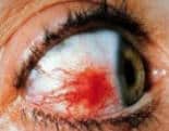
Communication with physicians managing
the patient’s systemic disease is crucial.
By Katrina Altersitz
Women are as much as 50 times more likely to suffer from
certain autoimmune diseases than men, and these diseases
often have ocular manifestations that ophthalmologists
should be aware of when examining their female patients,
experts say.
“Autoimmune diseases are the No. 1 example of the higher
risk” women carry for certain diseases due to sex, said
Janine A. Smith, MD, of the National Eye Institute. “There
are a lot of autoimmune diseases, and nearly every single
one affects women more than men.”
There is no definitively known reason for this disparity
between the sexes, she said.
Dr. Smith said Sjögren’s syndrome, with its symptoms of
dry eye and dry mouth, is a prime example of an autoimmune
disease seen more often in women. In addition, she said,
systemic lupus erythematosus, rheumatoid arthritis and
thyroid disease all disproportionately affect women, and all
can have ophthalmic manifestations.
“The immune system between men and women is different,”
Dr. Smith explained. “Even the way men respond to viruses
and women respond to infections, there are differences.”
One theory, she said, is that because estrogens are
naturally proinflammatory and androgens, specifically
testosterone, are considered anti-inflammatory, women are
more prone to developing autoimmune problems.
OSN spoke with Dr. Smith and other experts regarding the
ocular complications of autoimmune disorders, what can be
done to treat them and how to work with internal medicine
specialists and others in management of patients with these
diseases.
Lupus
Systemic lupus erythematosus, a collagen vascular
disease, affects women nine times as often as men. Typically
young, middle-aged women are affected, said C. Stephen
Foster, MD.
Lupus often causes dry eye, for which treatment is much
like that for Sjögren’s syndrome, but there are other
possible ocular complications as well. Usually, Dr. Foster
said, these complications are inflammatory.
“It might be something as relatively trivial as a rash, a
low grade subtle dermatitis on the lid, or something that is
directly relating to the eyeball itself with episodes of
superficial inflammation,” he said. “This superficial
inflammation may be affecting only the conjunctiva or, more
prominently, it will be episodic inflammation of the tissues
deeper to the conjunctiva like the episclera or sclera.”
Recurring episcleritis is a “trivial” but common ocular
complication of lupus, Dr. Foster said. While it rarely
requires aggressive therapy, episodes are often treated with
corticosteroid drops, which can have long-term consequences,
he explained.
“Eventually there is a price to pay, most particularly
with respect to development to cataract,” he said. “We
believe taking an oral nonsteroidal anti-inflammatory
medication is the most productive and appropriate way to
address that.”
Keratitis is another ocular manifestation of lupus.
Patients can develop peripheral keratitis or multifocal
superficial corneal inflammation, leaving small nebulae in
the superficial cornea stroma.
“Both of them are associated with light sensitivity and
photophobia. Both are important because they both carry with
them a little more prognostic significance than does
episcleritis or conjunctivitis,” Dr. Foster explained. “Both
are fairly disabling because of the photophobia.”

Kathryn Colby |
|
He said, again, corticosteroids are usually used for
treatment, but the long-term solution for keratitis is to
address the underlying lupus with systemic medication.
The systemic manifestations of lupus make it a
potentially life threatening disorder, said Kathryn Colby,
MD, PhD.
“The treatment is first identification of the systemic
disease associated with it,” she said. “Often, management of
the systemic disease will help with the ocular
manifestations.”
Unfortunately, Drs. Colby and Foster said, Plaquenil (hydroxychloroquine,
Sanofi Winthrop), the traditional systemic treatment for
lupus, can cause damage to the retina when used in high
dosage.
“There is a danger dose, which is measured in milligrams
per kilogram, so a smaller person would be more likely to be
at that danger dose using a regular dose of Plaquenil,” Dr.
Colby said. “It causes a retinopathy, basically dysfunction
of the retina cells.”
The site for some of the most serious ocular
complications of lupus is the retina, Dr. Foster said.
“Patients with lupus can develop an immune complex
vasculitis with the lodging of immune complexes forming in
the vasculature, both in the choroid and in the retinal
vasculature itself,” he explained.
This ophthalmic emergency can cause permanent vision
loss, but it can also be a prompt for systemic lupus testing
in an as yet undiagnosed patient or a sign of similar immune
complexes in other areas of the body.
“Ophthalmologists have a spectacular opportunity not only
to influence the situation but also to be lifesavers as well
if they make the diagnosis of lupus in the patient,” Dr.
Foster said. “The presence of lupus retinopathy with retinal
vasculitis or choroidopathy is a terrifically reliable
hallmark of the presence of these immune complexes lodging
in other areas as well, most particularly the brain and the
kidney. The likelihood of the patient dying from one of
those sites being affected by lupus nephropathy or lupus
affecting the central nervous system is very high in the
patient with lupus retinopathy who is not properly,
appropriately and aggressively treated.”
Rheumatoid arthritis
Rheumatoid arthritis, another collagen vascular disease,
is four times more common in women than men and presents
problems similar to those with lupus, including dry eye.
“You can also get scleritis and pretty severe
inflammatory disorders, things like melting of the
peripheral cornea,” Dr. Colby said. “There’s a lot of
overlap with lupus.”
Dr. Foster said the approach to treating dry eye or
secondary Sjögren’s in rheumatoid arthritis patients is much
like that in lupus, including the use of Restasis
(cyclosporine ophthalmic emulsion, Allergan) along with heat
and massage to improve the function of the meibomian glands.
In addition, he said, the ophthalmologist must be aware
of the patient’s systemic disease management to best treat
its ocular manifestations.
“See if there is any evidence of the underlying
rheumatoid disease being active, so to speak, even if it’s
not active in the joints,” Dr. Foster said. “Are there
markers in the blood that would indicate that chronic
low-grade inflammatory activity is afoot? If so, deal with
that systemically with a sprucing up of the systemic
medication program.”
Dr. Foster said that scleritis and peripheral keratitis
are all “absolutely dreadful” ocular manifestations of
rheumatoid disease that require systemic therapy, often
aggressive.
Dr. Colby added, “The important thing with the systemic
diseases is, the better the systemic diseases are managed,
the easier it is to manage the ophthalmic manifestations. I
can treat someone’s rheumatoid-associated dry eye all I
want, but if their systemic rheumatoid disease is not under
control it’s like sticking your finger in the dike.”
Scleroderma
Three times as many women as men experience scleroderma,
or progressive systemic sclerosis, a connective tissue
disease.

C. Stephen Foster |
|
“Scleroderma is a very different beast from lupus and
rheumatoid disease, with less known about it and less
discovered thus far in terms of a routinely highly effective
therapy,” Dr. Foster said. “Breakthroughs are being made,
and there are increasing levels of optimism among the
medical community with respect to being able to prevail over
some of the more relentless manifestations of scleroderma.”
Patients with scleroderma are likely to develop dry eye,
which is treated as in other autoimmune diseases. The
condition also causes shrinkage of areas of the skin,
including the conjunctiva, Dr. Foster said.
“Patients sometimes develop shrinkage of the conjunctiva
with a shortening of the inferior fornix,” he said. “It
probably has to do with laying down new collagen, perhaps
type 3 collagen or embryonic collagen. This contracts and
you get shrinkage.”
Choroidopathy and vascular occlusion can also occur.
“Just as in lupus, this is a wake-up call that the
patient’s scleroderma has taken a nasty turn,” Dr. Foster
said.
Unfortunately, he said, there is no treatment for
conjunctival shrinkage, and clinical trials for treatment of
vascular occlusions are “maddeningly difficult” because of
the small number of patients in any one center.
“One could speculate that the same approach that has been
employed in the scleroderma lung research, namely with high
dose cyclophosphamide therapy intravenously and other kinds
of aggressive immunosuppressive therapy, might be beneficial
in patients who are developing the scleroderma choroidopathy,
but frankly it’s not known,” Dr. Foster said.
Hyperthyroiditis
Hyperthyroiditis affects women seven times as often as
men. Its main ocular manifestation is Graves’ orbitopathy.
“That often is relatively mild and basically cosmetic,”
Dr. Foster said, “but it is graded and assigned different
grades, with severity and vision threat being greater the
higher the grade of the thyroid eye disease is.”
Dr. Colby explained that in Graves’ orbitopathy
infiltration of inflammatory cells within the extraocular
muscles causes proptosis.
“You can have exposure of the cornea, which can lead to
corneal ulcers. More seriously, if the muscles are very
infiltrated, “crowding” of the orbit can compress the optic
nerve of the eye, which can cause vision loss,” she said.
“Graves’ disease can also cause double vision because if the
muscles are very infiltrated they don’t move properly.”
Mild exposure can be treated with ointment or taping of
the lids, Dr. Colby said. More advanced disease may require
partial tarsorrhaphy. In extreme cases, orbital
decompression surgery may be called for.
Dr. Foster said, “As the space occupied by the
infiltrating cells, the proliferating fibroblasts, steadily
increases, there is only so much space in the orbit, and the
optic nerves can get increasingly compressed. That’s
something that is typically tackled surgically with
decompression surgery.”
Working with other physicians
Given that ocular manifestations of autoimmune disease
are related to the status of the systemic disease, Drs.
Foster and Colby said ophthalmologists must maintain
relationships with the patient’s ocular immunologist or
rheumatologist during treatment.
“With any of those diseases, the systemic management is
really key,” Dr. Colby said. “Often if the eye is out of
control, it means the body is out of control, even if the
patient is not having systemic symptoms.”
She suggested that ophthalmologists make sure to speak
clearly with collaborating physicians, using terms that the
other specialist will understand.
“It’s important to really be convinced of the concept
that appropriate systemic management will only make the
ophthalmologist’s life easier and make it easier to manage
patients’ problems,” Dr. Colby said.
“Communication is critical,” Dr. Foster agreed.
He said good communication will come into play when the
eye reveals deeper problems in the body. If this is the
case, it is the responsibility of the ophthalmologist to
demonstrate that to the rheumatologist and to ensure that
the patient receives proper systemic care.
“The patient, from a rheumatologist’s standpoint, may
still be in remission,” Dr. Foster said. “The
ophthalmologist is duty-bound, in my judgment, to convince
that rheumatologist that this is not so – that the eye,
although it may seem trivial, is extremely important and is
telling you, ‘Wake up, pay attention, increase the
aggressive therapy.’ If you don’t, you’re going to be sorry
6 months from now because the kidney or the lung is going to
be in trouble.”
For more information:
-
Janine A. Smith, MD, can be reached at the National
Eye Institute 31 Center Drive, MSC 2510, Bethesda,
MD, 20892; 301-496-9058; fax: 301-496-2297.
-
Kathryn Colby, MD, PhD, can be reached at the
Massachusetts Eye and Ear Infirmary, 243 Charles
St., Suite 808, Boston, MA 02114; 617-573-5537; fax:
617-573-3364. Dr. Colby has no direct financial
interest in the products discussed in this article,
nor is she a paid consultant for any companies
mentioned.
-
C. Stephen Foster, MD, can be reached at Ocular
Immunology and Uveitis Foundation, Massachusetts Eye
Research Surgery Institute, 5 Cambridge Center,
Eighth Floor, Cambridge, MA 02142; 617-742-6377;
fax: 617-227-1185.
-
Katrina Altersitz is an OSN Staff Writer who covers
all aspects of ophthalmology.
|