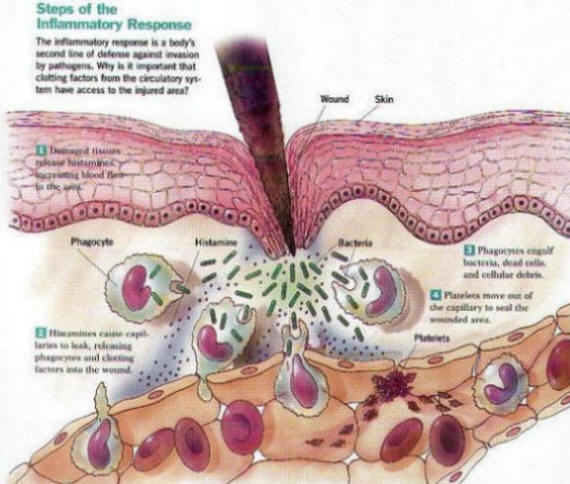
Macrophages and neutrophils that invade blood vessels produce many toxic molecules and contribute to damage of the blood vessels. In rheumatoid arthritis, reactive oxygen intermediate molecules and other toxic molecules are made by overproductive macrophages and neutrophils invading the joints. The toxic molecules contribute to inflammation, which is observed as warmth and swelling, and participate in damage to the joint.
MHC and Co-Stimulatory MoleculesMHC molecules are found on all cell surfaces and are an active part of the body's defense team. For example, when a virus infects a cell, a MHC molecule binds to a piece of a virus (antigen) and displays the antigen on the cell's surface. Cells that have the capability of displaying antigen with MHC are called antigen-presenting cells. Each MHC molecule that displays an antigen is recognized by a matching or compatible T-cell receptor. Thus, an antigen-presenting cell is able to communicate with a T cell about what may be occurring inside the cell.
Internet help for people in any locations Available contact us
| However, for the T cell to respond to a foreign antigen on the MHC, another molecule on the antigen-presenting cell must send a second signal to the T cell. A corresponding molecule on the surface of the T cells recognizes the second signal. These two secondary molecules of the antigen-presenting cell and the T cell are called co-stimulatory molecules. There are several different sets of co-stimulatory molecules that can participate in the interaction of antigen-presenting cell with a T cell. Once the MHC and the T-cell receptor interact, and the co-stimulatory molecules interact, there are several possible paths that the T cell may take. These include T cell activation, tolerance, or T cell death. The subsequent steps depend in part on which co-stimulatory molecules interact and how well they interact. Because these interactions are so critical to the response of the immune system, researchers are intensively studying them to find new therapies that could control or stop the immune system attack on self tissues and organs. Cells and molecules of the immune system protect the nose from attack by a virus. . An antigen-presenting cell (for example, a macrophage) with a foreign antigen on its MHC is recognized by a T-cell receptor. In addition, co-stimulatory molecules on the antigen-presenting cell and the T cell interact. Cytokines and ChemokinesOne way T cells can respond after the interaction of the MHC and the T-cell receptor, and the interaction of the co-stimulatory molecules, is to secrete cytokines and chemokines. Cytokines are proteins that may cause surrounding immune system cells to become activated, grow, or die. They also may influence non-immune system tissues. For example, some cytokines may contribute to the thickening of the skin that occurs in people with scleroderma.
|