| |
Guide to
treatment of autoimmune disease by alternatives please read our
e-book
See our services section for
help and contact information.
continued from the Brain Page of
Nervous System
Contents
neurons and Nerves
neurotransmitter
The Brain & Spinal Cord
Cranial Nerves
Peripheral Nervous System
Autonomic Nervous System
Senses:
Eye diagrams,
Hearing,
Smell,
Taste, Taste
& Tongue Sensation,
Balance
Memory ,
Memory types, Creation of Memory,
Higher Functions
Altered States
[Top]
Continued from Brain
 |
 |
The human brain can be divided into three
parts: the hindbrain, which has been inherited from the
reptiles; the limbic system, which was first emerged in
mammals; and the forebrain, which has its full development
in human. Different views of the human brain are shown in
Figure 03c and 04d. Tables 01 lists the functions of the
different parts of the human brain. The brain is separated
into two hemispheres. Apart from a single little organ --
the pineal gland in the centre base of the brain -- every
brain module is duplicated in each hemisphere. The left
brain is calculating, communicative and capable of
conceiving and executing complicated plans -- |
Figure 03c Human Brain
|
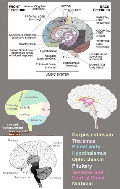
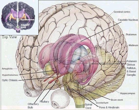
Figure 03d Human Brain
[view large image
c, d]
|
the reductionistic brain; while the right
one is considered as gentle, emotional and more at one with
the natural world -- the holistic brain. The
cerebral cortex is covered in a thin skin |
of deeply wrinkled grey tissue called the grey matter (densely
packed neurons for information processing). Each infold on the
surface is known as a sulcus, and each bulge is know as a gyrus.
While the white tissue inside are axons -- tentacles which reach out
to other cells (to relay information). The cortex can be broken down
into many functional regions, each containing thousands of cortical
columns (oriented perpendicular to the cortical surface). Columns
are typically about half a millimeter in diameter and contain about
one hundred thousand neurons. They are the units of cognition (the
mental process of acquiring knowledge by the use of reasoning,
intuition or perception). Table 02 below lists the location and
functions of the major components in the human brain.
| Structure |
Location |
Functions |
Hindbrain
(Reptilian
Brain) |
|
|
|
Medulla |
at the top of the spinal cord |
controls breathing, heart rate, and blood
pressure. |
|
Pons |
above the medulla |
regulates sensory information and facial
expressions. |
|
Cerebellum |
at the lower rear |
controls movement, coordination, balance,
muscle tone, and learning motor skills. |
|
Reticular Formation |
a network of nerves extends from the
medulla to the cerebrum |
monitors the general level of activity in
the hindbrain and maintains a state of arousal; essential
for the regulation of sleep and wakefulness. |
|
Midbrain (superior & inferior colliculus) |
above the pons between the hindbrain and
forebrain |
relays sensory information from the
spinal cord to the forebrain. |
|
Pineal
Gland |
on top of the midbrain behind the
thalamus (the
third eye¤ for fishes,
amphibians, reptiles, and some birds) |
involves in
circadian and circannual rhythms; possibly involves in
maturation of sex organs. |
Limbic
System
(Mammalian
Brain) |
|
|
|
Thalamus |
in the middle of the limbic system |
relays incoming information (except
smell) to the appropriate part of the brain for further
processing. |
|
Hypothalamus, Pituitary Gland |
beneath thalamus |
regulates basic biological drives,
hormonal levels, sexual behavior, and controls autonomic
functions such as hunger, thirst, and body temperature. |
|
Optic
Chiasm |
in front of the pituitary gland |
left-right optic nerves cross-over point. |
|
Septum |
adjacent to hypothalamus |
stimulates sexual pleasure |
|
Hippocampus |
within the temporal lobe |
mediates learning and memory formation. |
|
Amygdala |
in front of the hippocampus |
responsible for anxiety, emotion, and
fear |
|
Mammillary Body, Fornix |
linked to the hippocampus |
have a role in emotional behavior,
learning, and motivation. |
|
Caudate Nucleus, Putamen, Globus Pallidus,
Basal
Ganglia (Striatum) |
outside the thalamus |
involves in movement, emotions, planning
and in integrating sensory information |
|
Ventricles and Central Canal |
from tiny central canal within the spinal
cord to the enlarged hollows within the skull called
ventricles |
fills with cerebrospinal fluid for
mechanical protection. |
|
Cingulate Gyrus |
above corpus callosum |
concentrates attention on adverse
internal stimuli such as pain, contains the feeling of self. |
|
Corpus
Callosum |
under the cingulate gyrus |
is a bundle of nerve fibers linking the
cerebral hemispheres, involve in language learning. |
Forebrain
(Human
Brain) |
|
|
Frontal Lobe
(Conscious Brain) |
in front of the head |
controls voluntary movement, thinking,
and feeling. |
|
Prefrontal Cortex |
in front of the frontal lobe |
inhibits inappropriate actions, forms
plans and concepts, helps focus attention, and bestows
meaning to perceptions. |
|
Parietal Lobe |
in top rear of the head |
contains the primary somatosensory area
that manages skin sensation. |
|
Occipital Lobe |
in the back of the head |
contains the visual cortex to manage
vision. |
|
Temporal Lobe |
on each side of the head above the
temples |
contains the auditory cortex to manage
hearing and speech. |
Table 02 Human Brain
¤The parietal eye is not an eye in the traditional sense
in that it does not see images, but rather is a photosensitive organ
which only reacts to light and dark. The parietal eye is connected
to the pineal body and is used to trigger hormone production and
thermoregulation. It often shows up as either a dark spot or an
opalescent spot. Opsin proteins sensitive to blue and green light
has been identified in the cell.
 |
It is well known that the brain is an
electrochemical organ; a fully functioning brain can
generate as much as 20 watts of electrical power. Even
though this electrical power is very limited, it does occur
in very specific ways that are characteristic of the human
brain. Electrical activity emanating from the brain can be
displayed in the form of brainwaves. There are four
categories of these brainwaves, ranging from the most active
to the least active. Figure 03e is produced by an
EEG
(ElectroEncephaloGraph) chart recorder to show the different
kind of brainwave according to the different state of the
brain. These are all oscillating electrical voltages in the
brain, but they are very tiny voltages, just a few
millionths of a volt. Electrodes are placed on the outer
surface of the head to detect electrical changes in the
extracellular fluid of the brain in response to changes in
potential |
|
|
among large groups of neurons. The resulting
signals from the electrodes are amplified and recorded.
|
Brain waves originate from the cerebral cortex, but also reflect
activities in other parts of the brain that influence the cortex,
such as the reticular formation. Because the intensity of electrical
changes is directly related to the degree of neuronal activity,
brain waves vary markedly in amplitude and frequency between sleep
and wakefulness. Beta wave rhythms appear to be involved in higher
mental activity, including perception and consciousness. It seems to
be associated with consciousness, e.g., it disappears with general
anesthesia. Other waves that can be detected are Alpha, Theta, and
Delta. When the hemispheres or regions of the brain are producing a
wave synchronously, they are said to be coherent. Alpha waves are
generated in the Thalamus (the brain within the brain), while Theta
waves occur mainly in the parietal and temporal regions of the
cerebrum. The Alpha and Theta waves seem to be associated with
creative, insightful thought. When an artist or scientist has the
"aha" experience, there's a good chance he or she is in Alpha or
Theta. These two kinds of brain waves are also associated with
relaxation and, stronger immune systems. Therefore, many people try
to train themselves to enter such states through various biofeedback7
techniques (with varying degree of success). Delta Waves occur
during sleep. They originate from the cerebral cortex when it is not
being activated by the reticular formation. In slow-wave sleep, the
entire brain oscillates in a gentle rhythm quite unlike the
fragmented oscillations of normal consciousness. The neocortical
activity is often modulated by a rhythm of 40-80 Hz, called the
Gamma wave (not shown in Figure 03e). When there are strong gamma
oscillations in certain parts of the neocortex, human subjects do
better on learning and memory tasks.
[Top]
 |
 |
The spinal cord (Figure 04a) lies along the
middorsal line of the body. It has two main functions: (1)
it is the center for many reflex actions, and (2) it
provides a means of commu-nication between the brain and the
spinal nerves that leave the cord (Figure 04b). The white
matter of the cord is white because it contains myelinated
long fibers of interneurons that run to-gether in bundles
call tracts. These tracts connect the cord to the brain. The
dorsal ones are primarily ascending to the brain, while the
ventral tracts bring information down from the brain. The
inner portion of the |
|
|
cord is filled with a mass of nerve cell
bodies called gray matter. Each spinal nerve emerges from
the spinal cord as two short branches, the dorsal and the
ventral roots. These roots join just before the nerve leaves
the vertebral column. |
[Top]
 |
 |
The peripheral nervous system is outside the
CNS. It consists of the various nerves that connect
particular parts of the CNS with particular organs. Humans
have 12 pairs of cranial nerves and 31 pairs of spinal
nerves. Cranial nerves (Figure 05) are either sensory
nerves, motor nerves, or mixed nerves. All of them, except
the vagus nerve, control the head, the face, the neck, and
the shoulders. The vagus nerve controls the internal organs.
Table 03 lists the functions of the various cranial nerves.
All spinal nerves (Figure 06) are mixed nerves that take
impulses to and from the spinal cord. Table 04
|
|
|
|
describes the symptom of spinal cord injury
(SCI) with the particular spinal nerve(s).
|
Continue to next page the Cranial
Nerves |
World Wide Consultation by Internet
DHEA levels and
cognitive functions
SESAME SEED OIL
Facial cleaner
Quran
page |