| |
Complete
guide to alternative
treatment of autoimmune disease please read our
e-book
See our services section for
help and contact information. continued
from the Brain Page of Nervous System
Contents
neurons and Nerves
neurotransmitter
The Brain & Spinal Cord
Cranial Nerves
Peripheral Nervous System
Autonomic Nervous System
Senses: Eye
diagrams, Hearing, Smell,
Taste, Taste
& Tongue Sensation,
Balance
Memory ,
Memory types, Creation of Memory,
Higher Functions
Altered States
[Top]
Continued from Brain
The general receptors distribute all over the skin. They are usually
grouped together as sensation. The special receptors locate only at
certain part of the body in the head. Altogether, they are referred
to as the five senses. The followings present a further break down
into components, and functions.
Sight (see location of the various
components in Figure 09):
 |
- Sclera - This is the white of the eye. It is a tough
whitish sheath covering most of the eye (see Figure 11).
The inter-cellular space contains a kind of loose
proteins composed of collagen and elastic fibers, which
make the wall of the eyeball more pliant.
- Cornea - It is almost perfectly transparent at the
front of the sclera for the protection of the eye. Light
rays is refracted through this dome-shaped structure.
- Choroid - The soft layer inside the sclera. It has
many blood vessels that nourish the eye. It also absorbs
stray light.
|
|
|
- Ciliary body - The choroid becomes suspensory
ligaments near the opening. It holds the lens in place,
and controls the shape of the lens for near and far
vision.
|
- Iris - Further toward the opening, the choroid becomes a
thin, circular, muscular diaphragm known as the iris, which
regulates the size of a center hole (the pupil) and thus
controls the amount of light entering into the eye. The pupil
gets larger in dim light and smaller in bright light.
- Aqueous humor - This is the anterior cavity between the
cornea and the lens. It is filled with an alkaline, watery
solution secreted by the ciliary body. Normally, the solution
leaves the anterior cavity by way of tiny ducts that are located
where the iris meets the cornea. When a person has glaucoma,
these drainage ducts are blocked, and aqueous humor builds up.
The resulting pressure compresses arteries that serve the nerve
fibers of the retina. The nerve fibers begin to die due to lack
of nutrients, and the person becomes partially blind.
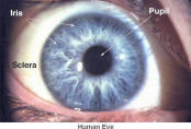
- Lens - It is a fat disk that completes the focusing of light
rays into a clear, sharp image on the retina. Actually, the
cornea provides about 80% of the focusing power. The lens is
stretched by the ciliary muscles to fine-tune the focus by
changing the shape, so it becomes fat for near objects, thin for
far ones.
- Vitreous humor - The posterior cavity behind the lens
contains a viscous, gelatinous material. It forms the bulk
inside the eyeball and, with the outer sclera, gives it
firmness. It also helps to refract light rays toward the retina.
 |
 |
- Retina - Inside each light-sensitive cell (rod or
cone) in the retina are up to 100 million molecules of
photopigment, each of which contains a smaller molecule
known as retinal (Figure 12a). When retinal receives
light energy, it changes shape by twisting around its
backbone. The altered retinal sets off a chain of
chemical reactions inside the cell, which triggers an
electro-chemical change in the cell membrane creating a
nerve impulse. The retinal returns to its original
configuration when the signal jumps across a synapse to
a bipolar cell. Curiously, in order to
|
|
|
|
reach the photoreceptors, incoming light
must first pass through all the other layers of cells in
the retina. There are five layers altogether (see Figure
12b). Starting from the outermost layer: |
- Pigment epithelium - Epithelial cells are the guards and
protectors of the organ. They cover the surface and
determine which substances are allowed to enter.
- Rods and cones - These are the photoreceptors. The rods
are responsible for night vision, and the cones for color
vision. The retina has as many as 150 million rods but only
1 million ganglionic cells and optic nerve fibers. This
means that there is considerable mixing of messages and a
certain amount of integration before nerve impulses are sent
to its final destination. The photoreceptors achieve the
remarkable sensitivity (of a millionfold difference in
luminance) with adjustment relative to the average
background. But if the overall background level of
illumination were to change drastically, as it does when we
enter a dark room, we are effectively blind for a few
minutes until the rod photoreceptors have adapted to this
level of reduced intensity. Vision is most acute in the
fovea centralis (see Figure 09), where
there are only cone cells. Clear vision is possible only
when the fovea inspects a scene. This provides a very
restricted window of clarity. Thus in order to obtain a
clear picture, the eyes have to dart about frenetically and
automatically under the direction of the brain.
- Bipolar cells - Nerve signals from the rods and cones
pass inward to this layer. The bipolar cells together with
the horizontal and amacrine cells form the network of
pre-processing nerve cells. The network helps to simplify,
and code information before it reaches the optic nerve.
- Ganglionic cell layer - It receives input from the
pre-processing network, and send output to the brain via the
axons of the ganglion cells.
- Optic nerve - It is made up of nerve fibers, which come
from across the whole retina. They pass in front of the
retina, forming the optic nerve, which turns to pierce the
layers of the eye. The signals eventually end up in the
occipital lobe to form an image.
 |
- Optic Pathway - As shown in Figure 13, information
about the left visual world is transmitted to the right
side of the brain and vice versa. As the visual fields
of the eyes overlap in the front, this division is
achieved by sorting retinal ganglion cell axons
according to whether they look at the left (dotted line)
or the right (solid line) visual field. So some axons
from the right eye go to the right side of the brain and
others to the left. The sorting occurs in the optic
chiasm. The retinal axons proceed to the lateral
geniculate nuclei where the first synapses are fromed
with the visual neurons in the brains.
|
|
|
|
|
World Wide Consultation by Internet
DHEA levels and
cognitive functions
SESAME SEED OIL
Facial cleaner
Quran
page |