Neurology Anatomy Physiology
December 14, 2021
Contents
neurons and Nervesneurotransmitter
The Brain & Spinal Cord
Cranial Nerves
Peripheral Nervous System
Autonomic Nervous System
Senses: Eye diagrams, Hearing, Smell, Taste, Taste & Tongue Sensation, Balance
Memory , Memory types, Creation of Memory
Higher Functions
Altered States
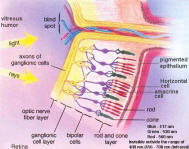
Figure 12a Retinal
[view large image]
Figure 12b Retina
[view large image]
reach the photoreceptors, incoming light must first pass through all the other layers of cells in the retina. There are five layers altogether (see Figure 12b). Starting from the outermost layer:
- Pigment epithelium - Epithelial cells are the guards and protectors of the organ. They cover the surface and determine which substances are allowed to enter.
- Rods and cones - These are the photoreceptors. The rods are responsible for night vision, and the cones for color vision. The retina has as many as 150 million rods but only 1 million ganglionic cells and optic nerve fibers. This means that there is considerable mixing of messages and a certain amount of integration before nerve impulses are sent to its final destination. The photoreceptors achieve the remarkable sensitivity (of a millionfold difference in luminance) with adjustment relative to the average background. But if the overall background level of illumination were to change drastically, as it does when we enter a dark room, we are effectively blind for a few minutes until the rod photoreceptors have adapted to this level of reduced intensity. Vision is most acute in the fovea centralis (see Figure 09), where there are only cone cells. Clear vision is possible only when the fovea inspects a scene. This provides a very restricted window of clarity. Thus in order to obtain a clear picture, the eyes have to dart about frenetically and automatically under the direction of the brain.
- Bipolar cells - Nerve signals from the rods and cones pass inward to this layer. The bipolar cells together with the horizontal and amacrine cells form the network of pre-processing nerve cells. The network helps to simplify, and code information before it reaches the optic nerve.
- Ganglionic cell layer - It receives input from the pre-processing network, and send output to the brain via the axons of the ganglion cells.
- Optic nerve - It is made up of nerve fibers, which come from across the whole retina. They pass in front of the retina, forming the optic nerve, which turns to pierce the layers of the eye. The signals eventually end up in the occipital lobe to form an image.
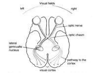
Figure 13 Optic Pathway
[view large image]
Powered by cidpusa.org