Neurology Anatomy Physiology
Aprial 24, 2020
Contents
neurons and Nervesneurotransmitter
The Brain & Spinal Cord
Cranial Nerves
Peripheral Nervous System
Autonomic Nervous System
Senses: Eye diagrams, Hearing,Smell,Taste, Taste & Tongue Sensation,Balance
Memory , Memory types, Creation of Memory,
Higher Functions
Altered States
| Cranial Nerve | CN# | Brain Region | Major Functions |
|---|---|---|---|
| Terminal§ | 0 | Near the olfactory | Reception of pheromone for sex |
| Olfactory | 1 | Cerebral Cortex | Smell |
| Optic | 2 | Limbic System | Vision |
| Oculomotor | 3 | Midbrain | Eyelid & eyeball movement; pupil dilation |
| Trochlear | 4 | Pons | Control downward & lateral eye movement |
| Trigeminal | 5 | " | Chewing; sensation of face & mouth |
| Abducens | 6 | " | Control lateral eye movement |
| Facial | 7 | " | Control most facial expressions; secretion of tears & saliva; taste; ear sensation |
| Auditory | 8 | Medulla | Hearing; balance |
| Glossopharyngeal | 9 | " | Taste; swallowing; sensation from tongue, tonsil, pharynx, carotid blood pressure |
| Vagus | 10 | " | Sensory, motor and autonomic functions of viscera - glands, digestion, heart rate, breathing rate, aortic blood pressure |
| Spinal Accessory | 11 | " | Controls muscles used in head movement |
| Hypoglossal | 12 | " | Controls tongue movements |
Table 03 Functions of Cranial Nerves
§ The exact function of the terminal nerve in human is still under investigation, which is hampered by its small size and proximity to the olfactory nerve. For mouse and other animals at least, it is connected to thevomeronasal organ (vestige in human), which leads to a pathway for controlling sexual arousal.
| Spinal Nerve(s) | Innervated Body Part(s) | Symptom(s) of SCI |
|---|---|---|
| C1 | Head and Neck | Quadriplegia |
| C2-C4 | Diaphragm | Breathing problem |
| C5 | Deltoids, biceps | No control at wrist or hand |
| C6 | Wrist extenders | No hand function |
| C7-T1 | Triceps, hand | dexterity problems with hand and fingers |
| T2-T8 | Chest muscles | Paraplegia, poor trunk control |
| T9-T12 | Abdominal muscles | Paraplegia |
| Lumbar and Sacral | Leg muscles, bowel, bladder, sexual organs | Decreasing control of hip flexors and legs, dysfunction of bowel, bladder, and sex |
Table 04 Symptom(s) of Spinal Cord Injury
Note: Other effects of SCI may include low blood pressure, inability to regulate blood pressure effectively, reduced control of body temperature, inability to sweat below the level of injury, and chronic pain.[Top]
Autonomic Nervous System
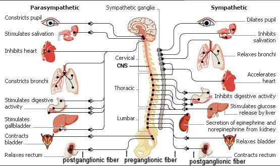
One division of the autonomic nervous system, called the sympathetic nervous system, dominates in times of stress. It controls the "fight or flight" reaction, increasing blood pressure, heart rate, breathing rate, and blood flow to the muscles. Another division, called the parasympathetic nervous system, has the opposite effect. It conserves energy by slowing the heartbeat and breathing rate, and by promoting digestion and elimination (of waste). Most glands, smooth muscles, and cardiac muscles constantly get inputs from both the sympathetic and parasympathetic systems. The CNS controls the activity by varying the ratio of the signals. Depending on which motor neurons are selected by the CNS, the net effect of the arriving signals
Figure 08 ANS Front View [view large image] | Figure 08 is the front view of a more detailed ANS anatomy. |
![[view large image]](images/I10-51-ANS2.jpg)
[Top]
Continue to Ganglia of autonomic page
alternatives treatment of autoimmune disease read our
e-book
Powered by cidpusa.org