Neurology Anatomy Physiology
December 14, 2021
Autonomic Nerve Ganglia
Nervous System
continued from the Brain Page of Nervous System
Contents
neurons and Nervesneurotransmitter
The Brain & Spinal Cord
Cranial Nerves
Peripheral Nervous System
Autonomic Nervous System
Senses: Eye diagrams, Hearing,Smell,Taste, Taste & Tongue Sensation,Balance
Memory , Memory types, Creation of Memory,
Higher Functions
Altered States
Figure 07 ANS Side View [view large image]
will either stimulate or inhibit the organ. Figure 07 shows the various organs and actions, which are related to the two different divisions. Motor fibers that govern involuntary responses, do not lead directly to the organs they innervate. Instead, they make their trips in two stages. The first set of fibers leads from the CNS to ganglia (which are collections of nerve cell bodies) that lie outside![[view large image]](images/I10-51-ANS.jpg)
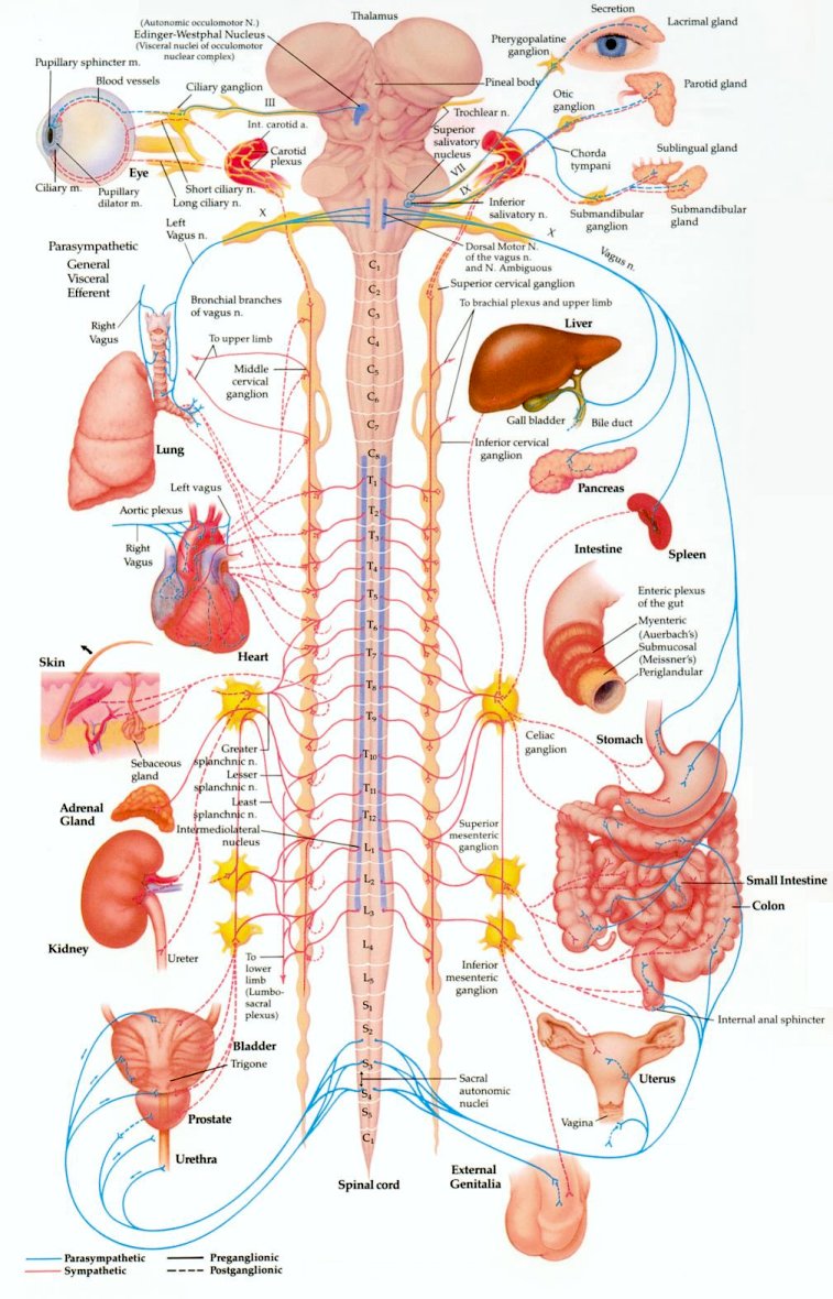
the CNS (the preganglionic fibers). At the ganglia the fibers form synaptic junctions with the dendrites of as many as twenty different cell bodies. The axons of these cell bodies form a second set of fibers, the postganglionic fibers. It is these postganglionic fibers that lead to the organs.
The chief ganglia involved in the autonomic nervous system form two lines running down either side of the spinal column. They are outside the bony vertebrae. These two lines of ganglia outside the column resemble a pair of long beaded cords. At the lower end, the two cords join and finish in a single central stretch. These lines of ganglia are sometimes called the sympathetic trunks (used by the sympathetic nervous system). Not all ganglia are located in the sympathetic trunks. Some are not; and it is possible for a preganglionic fiber to go right through, making no synaptic junction there at all, joining instead with ganglia located in front of the vertebrae. For the parasympathetic nervous system, some of the ganglia separating the preganglionic fibers from the postganglionic fibers are actually located within the organ the nerve is servicing. In that case, the preganglionic fiber runs almost the full length of the total track, whereas the postganglionic fiber is at most just a few millimeters long.
The splanchnic nerves, which originate from some of the thoracic nerves, have their preganglionic fibers ending in a mass of ganglia lying just behind the stomach. It represents the largest mass of nerve cells that is not within the CNS and is sometimes called the "abdominal brain". It is a vital spot to be protected during boxing.
Figure 08 ANS Front View [view large image]
Figure 08 is the front view of a more detailed ANS anatomy.Powered by cidpusa.org