|
;Global Health Help & information Welcome to cidpusa.org |
||||||||
| Guide to natural treatment of all diseases Flame within E-book. | ||||||||
|
IgA Nephropathyby Franco Ferrario and Maria Pia Rastaldi
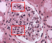
The characteristic and diagnostic lesion of idiopathic IgA nephropathy (IgA GN, Berger's disease) is represented by glomerular deposition of IgA, as a unique or predominant positive immunoglobulin. Despite the relatively uniform aspect of the immunohistological pattern, the morphological lesions observed by light microscopy are extremely variable. This variability of glomerular and interstitial alterations has frequently created some difficulties in histological classification. Glomeruli can show very mild lesions, without mesangial matrix expansion or mesangial cell proliferation. In other cases, mesangial matrix enlargement is conspicuous and predominates over a relatively mild mesangial cell proliferation. Most cases are characterized by focal and segmental mesangial proliferation (Fig. 1).
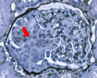
In approximately 20% of cases, mesangial matrix expansion and mesangial cell proliferation are particularly severe, with a morphological pattern that looks like idiopathic m embranoproliferative glomerulonephritis. Independent of the entity of mesangial damage, the presence of a necrotizing lesion has been described by many authors. The segmental tuft necrosis can be the only lesion highlighted, possible expression of an early phase of capillary damage. In most cases, the necrotic lesion is associated with an area of cellular extracapillary proliferation. By silver staining the necrotic damage is clearly evident, with segmental destruction of the glomerular basement membrane and associated extracapillary reaction (Fig. 2).
By monoclonal antibodies directed against leukocytes it has been possible to demonstrate the predominant presence of monocyte-macrophages (CD68), both in the areas of necrosis and in the segmental extracapillary reaction. In these necrotizing extracapillary forms, it is very common to find interstitial infiltrates with predominant periglomerular localization, adjacent to the areas of intraglomerular lesions. By silver staining a possible rupture of the Bowmans capsule is clearly demonstrated, making it difficult to discriminate between intra and extraglomerular lesions (Fig. 3). For this reason, many researchers believe that these forms should be treated with steroids and cyclophosphamide pulses. In rare cases it is possible to find a diffuse extracapillary glomerulonephritis with circumferential cellular crescents in more than 60-70% of glomeruli, clinically characterized by rapidly progressive renal failure. Marker of poor prognosis, diffuse interstitial infiltrates are commonly found in IgA GN. Though the diagnostic “marker” of IgA GN is represented by a predominant glomerular deposition of IgA, the immunohistological pattern is variable. An intense IgA deposition in the mesangial stalk is more common pattern (Fig. 5). | ||||||||
|
|
||||||||


